The following is a summary of the research article, Cortical thickness, volume and surface area in patients with bipolar disorder types I and II.
What is the topic?
Bipolar disorder (BD) is a chronic disorder that can be associated with disability, untimely death and cognitive difficulties. Previous studies have shown that there are structural changes in the outer layer of the brain (this layer is known as the ‘cortex’), such that there is lower cortical volume in patients with BD in certain areas of the brain.
BD is actually not one condition; rather, people can have BDI or BDII which differ in their symptoms. Most people think of BDII as a milder version of BDI. However, not a lot is known about what is different in the brains of individuals with BDI as compared to those with BDII, because researchers have either combined these two conditions together, or studies have been too small to determine differences.
What was the hypothesis?
Given that there are similarities and differences in symptoms in BDI vs. BDII, the researchers hypothesized that there would be common as well as unique findings between the conditions when they looked at cortical volume, thickness and surface area.
What did the researchers hope to learn?
The researchers examined the brains of people with BDI, BDII as compared to people without BD (controls). The hope was to analyze volume, thickness and surface area of the cortex (the outer layer of the brain) of people with BD to see if these parameters relate to patient symptoms.
Who was studied?
81 patients with BDI, 59 with BDII and 85 control patients (with no BD) participated, making this a large study. Participant characteristics e.g. age, smoking history, medical history etc were carefully noted, as they could impact the findings.
How was the study conducted?
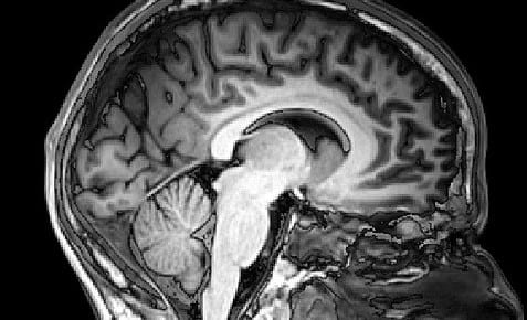
All participants underwent magnetic resonance imaging (MRI) to detect volume, thickness and surface area of the cortex. Images were collected and analyzed by the researchers.
What did the researchers find?
All patients with BD (irrespective of BDI or BDII) had aberrations in their cortices as compared to control patients. The interesting difference was that patients with BDI had more severe cortical abnormalities as compared to those with BDII. Lithium (a drug used to treat BD) and anti-epileptic drugs had an effect on the structure of the cortex.
What were the limitations of the study?
The researchers analyzed patients only at one point in time. So, it is difficult to say whether the structural changes were always present, or whether they developed over time.
What do the results mean for you?
This study reveals differences between BDI and BDII that were not yet known. It could be that MRI data could be used in the future to accurately diagnose BD. Eventually, drugs specific for BDI and BDII may also be developed.
This summary was written by Sloka Iyengar, PhD- a neuroscientist and science writer based in New York (September 2016). You can find more about her at www.slokaiyengar.com.